Contributed by: Healthians Team
Introduction
Did you know, According to statistics, more than 2 lakh women in India were expected to have a breast cancer diagnosis, and more than 76,000 fatalities were anticipated?
Breast tissue contains aberrant cells that begin to form and expand uncontrolled to develop breast cancer. Because the prognosis depends on the size and stage of the malignancy, an early detection is essential.
Even if you haven’t begun receiving mammograms on a regular basis, you should get to know your breasts so that you can see any changes and let your doctor know about them.
According to breast cancer studies from Johns Hopkins Medical Center, at least 40% of breast cancers are initially discovered by women who feel a lump while performing a self-exam.
Various diagnostic procedures aid in the diagnosis and early detection of breast cancer.
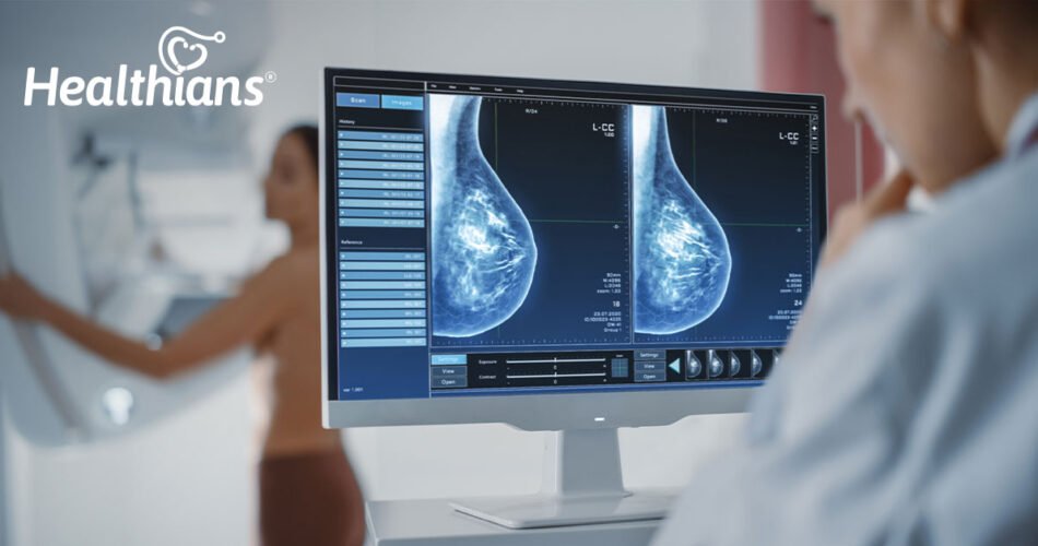
Image tests for Breast cancer
Imaging studies examine breast tissue using a variety of tools or technologies. There are several imaging examinations.
Mammograms are advised every year for women over 45, although screenings can start as early as 40.
An X-ray called a mammogram only captures images of the breasts—these pictures aid medical professionals in spotting breast anomalies such as lumps that could be cancerous.
Remember that even though you might require more testing if your mammography reveals an anomaly, breast cancer isn’t always the cause.
An ultrasound test creates pictures of the interior of your body using sound waves. Your doctor can ask for an ultrasound if your mammogram shows a mass.
If you have a breast lump that is clearly visible, your doctor could also recommend doing an ultrasound. An ultrasound can assist in establishing whether a lump or tumour is solid or packed with fluid. A benign cyst that is not malignant is frequently seen in a fluid-filled mass. This isn’t always the case, though.
Depending on how the ultrasound picture appears, certain tumours may be a mixture of fluid and solid, which is normally benign but may need immediate follow-up imaging or a sample.
Biopsies for Breast Cancer
In order to identify whether a lump or growth is malignant or benign, a biopsy involves removing a sample of tissue. Typically, this is an outpatient surgery.
Depending on the size and location of the tumour, a breast biopsy can be done in a number of different methods. An operation or needle biopsy may be performed by a surgeon or radiologist if the tumour is tiny and not suspicious.
A sample piece of tissue is taken from your breast by the doctor doing the surgery. Depending on your doctor’s advice, you may choose to perform this with or without imaging guidance.
In some cases, a surgical biopsy may be required. This removes all or certain parts of the lump. The surgeon may also remove any enlarged lymph nodes.
Types of biopsies
Biopsy with a fine-needle aspirator
When the lump is solid, this form of biopsy is used. A little bit of tissue is removed by the doctor using a fine needle for pathology analysis. When a cyst is detected, the doctor may want to inspect it to make sure there is no malignancy present.
Core needle biopsy
During this process, a sample of tissue up to the size of a pen is extracted using a bigger needle and tube. Feel, ultrasound or mammography are used to guide the needle. A mammography-guided biopsy will be performed if a woman has a finding that can only be spotted by a mammogram. A stereotactic breast biopsy is another name for this procedure.
Surgical biopsy
In this form of biopsy, the mass is completely removed (lumpectomy), partially removed (incisional biopsy), or neither (excisional biopsy, broad local excision). Before performing surgery, the surgeon may utilise a technique called wire localization to chart a path to the tumour if it is tiny or difficult to find by touch. A mammogram or ultrasound guidance can be used to implant a wire.
Test for sentinel nodes
A biopsy from a lymph node, where cancer is most likely to spread first. For breast cancer, a sentinel node biopsy is often performed on lymph nodes in the axilla or armpit area.
This test is performed to identify whether cancer exists in the lymph nodes on the breast’s cancerous side.
Image-guided biopsy
Using imaging methods like ultrasound, mammography, or MRI, a clinician can produce a real-time image of a worrisome spot that is difficult to detect or feel through your skin for an image-guided biopsy. This image will be used by your doctor to pinpoint the ideal area for a needle.
Breast cancer stage-specific tests
The next step after receiving a breast cancer diagnosis is to determine your stage. Your doctor chooses the appropriate course of therapy based on the stage. The tumour’s size, location, and whether it has progressed outside of your breast to adjacent lymph nodes and other organs are all factors that influence the stage. After receiving a breast cancer diagnosis, the next step of staging is knowing the rate of growth and the possibility of staging element is the growth rate at which the growth will spread.
In order to assess the severity of cancer and aid in the diagnosis, your doctor may also employ any of the tests listed below.
Bone scan
Bones can become infected with breast cancer. Using a radionuclide tracer, a bone scan enables your doctor to examine your bones for signs of abnormalities.
CT scan
Another kind of X-ray that uses iodine contrast to get precise pictures of your organs is the CT scan. To determine whether cancer has progressed to organs other than the breast, such as the chest, lungs, or stomach region, your doctor may use a CT scan.
MRI scan
This imaging technique, while not often used for cancer screening, is useful for staging breast cancer.
Digital pictures of various body sections are produced during an MRI. It may assist your doctor in determining whether cancer has progressed to your brain, spinal cord, or other organs.
PET Scan
A PET scan is an uncommon test. A dye is injected into your vein by your doctor. A specialised camera creates 3-D photos of the interior of your body as the dye passes through it. This aids in locating cancers for your doctor.
Genetic testing for breast cancer
Another risk factor for breast cancer is genetics. According to medical experts, heredity is believed to be a factor in 5% to 10% of breast cancer cases. Consult a genetic counsellor and bring up obtaining a genetic test if you’re worried about your chance of developing breast cancer.
These tests involve drawing blood, collecting saliva, or scraping the cheek. There are steps you may take to avoid breast cancer if you determine that you are at high risk.
You could wish to have regular and early breast cancer tests, alter your drinking and exercise habits, get a mastectomy as preventive surgery, and more.
Final thoughts
Follow up with additional diagnostic testing if your mammogram or clinical exam reveals any anomalies. Although breast cancer is curable, if it is not found early enough, it can be incurable.
For further information about yearly screening, speak with your doctor, especially if you have a personal or family history of breast cancer.
Book The Full Body Good Health Test Today!


